Detection of PTEN by in situ molecular hybridization and immunohistochemical SP method

Detection of PTEN by in situ molecular hybridization and immunohistochemical SP method
【Abstract Objective:
In situ hybridization and immunohistochemical sP assay were used to detect the expression of PTEN mRNA and its protein in HCC.
Methods: 56 cases of hepatocellular carcinoma and 17 cases of cirrhosis were surgically removed. The specimens were fixed with 40g/L formalin, serial sections of 4 m thick, HE staining and pathological diagnosis.
Results: The positive expression rates of PTEN mRNA~ protein in HCC tissues were 62.5% and 71.4%, respectively. 17 cases of cirrhosis
The positive expression rates in the tissues were 88.2% and 94.1%, respectively. The positive rates in 21 adjacent tissues were 90.5% and 100%, respectively. There was a significant difference in the expression of PTEN mRNA and protein between HCC and cirrhotic tissues and paracancerous liver tissues (P<005).
Conclusion: There is a high proportion of PTEN mRNA and protein expression in HCC tissues, and the positive expression rate of PTEN mRNA and protein is the lowest in Hcc tissues with the highest degree of malignancy.
The PTEN (phosphatase and tensin homologuedeleted on chromosome ten) gene is a novel tumor suppressor gene discovered in 1997. It has mutations and deletions in many tumors and is closely related to tumorigenesis. PTEN has dual activities of lipid phosphatase and protein phosphatase, and is the first tumor suppressor gene with lipid phosphatase activity discovered so far. PTEN protein phosphatase activity has the effect of inhibiting cell invasion; PTEN lipid phosphatase activity has the effect of inhibiting cell cycle progression and inducing apoptosis. Hepatocellular carcinoma (HCC) is one of the common malignant tumors. The occurrence of HCC is a very complex and multi-step process in which there may be multiple changes in tumor suppressor genes. Therefore, the in-depth study of tumor suppressor genes is of great significance for the clarification of the pathogenesis of HCC and for the prevention and treatment of HCC.

1 research objects and methods
From 2007 to 2008, 56 cases of hepatocellular carcinoma and 17 cases of cirrhosis were surgically removed from the pathology department of Hunan Provincial Cancer Hospital. They were fixed with 40g/L formalin, 4"m thick serial sections, HE staining, pathological diagnosis. It was confirmed that the HCc classification was divided into 7 cases of high differentiation (Class I), 28 cases of moderate differentiation (Class II), 21 cases of poor differentiation (Class II) according to WHO criteria. 21 cases with adjacent liver tissues, including cirrhosis 11 cases, 5 cases of chronic active hepatitis, 5 cases of chronic persistent hepatitis. In situ hybridization: 1) section conventional dewaxing to water, 3% HO blocked endogenous peroxidase, room temperature for 10 min; 2) distilled water wash 3X5min. 3) 3% citric acid freshly diluted pepsin, digested at 37~C for 30 min, fully exposed mRNA nucleic acid fragments. 4) 0.5 M, pH 7.4 PBS buffer was shaken 3 × 5 arin. 5) Drop resistance Broken solution, 40C for 2h to prevent non-specific hybridization. Pour off, do not wash. 6) Add pre-hybrid solution, 40~C for 2h. Drain, do not wash. 7) Add hybrid solution, 42E hybrid overnight 8) 2×SSC buffer at 30~37°C water temperature for 2×15min. 9) Add blocking solution, 37E30min. Dip, do not wash. lO) Add anti-digoxigenin antibody base Phosphatase complex, 37 ° C for 60 min. 11) o. 5M, pH 7.4 PBS buffer for 4 x 5 min. 12) ~ o color development liquid color, 1% hydrochloric acid alcohol differentiation, sequential alcohol, xylene dehydration transparent, Neutral gum seal, positive liver tissue as a positive control, pre-hybrid solution instead of hybridization as a negative control. Immunohistochemical SP method: 1) section conventional dewaxing to water, 30mL · L methanol - HO liquid closed endogenous Peroxidase, 30 arin at room temperature. 2) Water rinse, 0.01 M, pH 7.4 in PBS buffer for 10 min. 3) Microwave antigen retrieval: sliced ​​in citrate containing 0.01 M, pH 6.0 In the horizontal tank of the buffer, the plastic film covered with the vent hole is heated at high temperature for 1-2 min. After the liquid is boiled, it is adjusted to the low level and heated for 8 min. At room temperature, the slice is naturally cooled. 4) 0. 01M, pH 7.4 PBS buffer was shaken 3×5rain. 5) Normal goat serum was blocked at room temperature for 30 min. Pour off, do not wash. 6) Add 1:100 rabbit anti-PTEN polyclonal antibody at 4 ° C overnight. ) 0.01 M, pH 7.4 in PBS buffer for 3 x 5 min. 8) Add biotinylated goat anti-rabbit IgG, 37 ° C-labeled chain enzyme White pigment, 37 ° C for 30 min. 11) o.01M, pH 7.4 PBS buffer for 3x5min. 12) DAB color development, hematoxylin counterstaining, l% hydrochloric acid alcohol differentiation, sequential alcohol, xylene dehydration transparent, medium Sexual gum seals, observed under light microscope. Normal liver tissue
As a positive control, 0.01 M, pH 7.4 PBS was used instead of the primary antibody as a negative control. Judgment and statistical analysis: Apparent light blue particles (in situ molecular hybridization) and brownish yellow particles (immuno-organized into PTEN-positive cells) in the cytoplasm. According to the ratio of positive cells to all cells in liver tissue Divided into: negative: no positive tumor cells, positive (+): positive cells / J, 25% positive (++): positive cells accounted for 25% ~ 5O% positive (+++): positive cells greater than 5 & / o. Statistical analysis Statistical analysis was performed using Sf 10.O statistical software. The two-sample composition ratio test was used. Statistically significant é™› difference ~ gNP

2 results
2.1 in situ hybridization PTEN
The mRNA was localized to the cytoplasm, and the positive reaction product was light blue particles, and the PTEN mRNA positive cells were diffusely distributed. The expression of PTEN mRNA was positive in 3 normal liver tissues; the positive rate of PTEN mRNA expression was 88.2% (15/17) in 17 cases of cirrhosis, see Figure 1; PTEN mRNA in 21 cases of adjacent liver tissue The positive expression was 3, g~j90.5% (19/21), as shown in Figure 2; and the positive rate of PTEN mRNA expression in 56 HCC tissues was 62.5% (35/56), as shown in Figure 3. The expression of PTEN mRNA in paracancerous liver tissues and cirrhosis tissues was significantly higher than that in HCC tissues (P<0.05, Table 1). In 56 cases of HCC tissues, the expression of PTEN mRNA of grade I and II was significantly higher than that of grade III (P

2.2 Immunohistochemistry results
The PTEN protein is mainly localized in the cytoplasm, and the positive reaction product is brown or brownish yellow particles, which is diffusely distributed. The expression of PTEN protein in 3 normal liver tissues was strongly positive (+++); the positive rate of PTEN protein expression in cirrhotic tissues was 94.1% (16/17), mainly strong positive expression; 21 cases adjacent to cancer Liver tissues were strongly positive: the positive rate of PTEN protein expression in 56 HCC tissues was 71.4% (40/56) (Table 3), and the immune response was different. In some specimens, the positive signal was weak. . The positive expression rate of PTEN was significantly higher in paracancerous tissues and cirrhotic tissues than in HCC tissues (P<0.O1). In 6 HCC tissues, the positive rate of PTEN protein expression of grade I and II was significantly higher than that of grade III (P <0.05, Table 4), but there was no significant difference between grade I and II. The positive expression intensity of #hi and II PTEN proteins was significantly higher than that of grade III, mostly ++~+++, while in grade III tissues, PTEN staining was mostly weakly positive or negative.

3 Discussion
In this study, in situ hybridization and immunohistochemistry were used to detect PTEN mRNA and PTEN protein in 56 HeC tissues, 17 cirrhotic tissues and 3 normal liver tissues. PTEN mRNA and PTEN protein were positively expressed in all 3 normal liver tissues. In 17 cases of cirrhosis, PTEN mRNA and PTEN protein expression were positively expressed at different intensities, and the positive rates were 88.2% (15/17) and 94.1% (16/l7), respectively. In 56 cases of HCC, the positive expression rates of PTEN mRNA and PTEN protein were 62.5% and 71.4‰, respectively. In 21 cases of adjacent tissues, the positive expression rates of PTEN mRNA and PTEN protein were 90.5%. 19/21) 30min. 9) Rinse 3X5arin in 0.01 M, pH 7.4 PBS buffer. 10) Add horseradish enzyme and 100%. Statistical analysis showed that the expression of PTEN mRNA and PTEN protein in paracancerous and cirrhotic tissues was significantly different from that in HCC tissues (P<0.05). This indicates that PTEN mRNA and PTEN protein are absent in the development of HCC, suggesting that it may play a role in the development of HCC. So far, studies on the PTEN gene in HCC tissues and cells have been limited to the study of gene mutations and deletions, and the expression of PTEN mRNA and its protein in liver cancer tissues has rarely been reported. The results of this study indicate that the loss of expression of PTEN at mRNA and protein levels is highly promising in HCC tissues with high malignancy, and it is possible that one of the main ways in which ~_PTEN gene is inactivated in HCC. Therefore, profoundly elucidating the role of PTEN gene in normal human tissues and its mechanism of action, it is of great significance to determine the pathogenesis of hepatocellular carcinoma and adopt corresponding effective gene therapy methods.

4 Conclusion
Both PTEN mRNA and its protein are mainly localized in the cytoplasm. Their expression in adjacent tissues and cirrhosis tissues was significantly higher than that in HCC tissues<0. O5). The expression of PTEN mRNA and its protein in HCC tissues was positively correlated with the degree of HHCC differentiation. The higher the degree of differentiation of HCC, the higher the expression rate of PTEN mRNA and its protein. It is suggested that the PTEN gene may play a role in the development of liver cancer.
(Modern medicine Wang Jin)

Collagen is a triple helical protein which can be considered as the bio-glue inside our body; in fact, animal glue can be obtained by boiling the animal skin.  Collagen, a major component of connective tissues, exits in the extracellular space of these tissues which are the key reinforcing and bonding materials for all tissues and organs throughout our body, forming rigid structures as such bone, semi-rigid tissues such as cartilage, or soft tissues such as muscle, tendon, skin, ligaments, and cell membranes, etc.  There are different forms (fibrillar and non- fibrillar) and types of collagens in the body; Type 1 being the major type constitutes over 90% in our body and is the major component in skin, tendon, vascular ligature, organs, bone (main component of the organic part of bone).   Because collagen is an essential building material of all tissues and organs, it has many medical uses, such as in cardiac (hear) applications, cosmetic surgery, bone grafts, tissue regeneration, reconstructive surgical uses, and wound healing care.   


Collagen is created inside fibroblast cells, and this process is needed to support the creation and repair of the body`s connective tissues.  However, the biological process starts to breakdown when we are aging, normally after we reach the age of late 20s or early 30s.  Because collagen from natural sources such as animal, fish scales or plant contain essentially the same amino acid compositions (glycine, proline, alanine, hydroxyproline, glutamic acid, arginine, aspartic acid, serine, lysine, leucine, valine, threonine, phenylalanine, isoleucine, etc.) as human collagen, supplement the body with the natural collagen, either by dermal application or through oral ingestion, can help rejuvenate collagen creation process to support the repairing of aging connective tissues in our body, particularly those in our skin, and to reverse or slow down the aging process for a more youthful appearance.

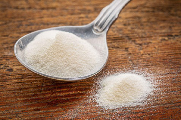
 CollagenCollagen

Collagen

Hydrolyzed Collagen,Fish Collagen,Collagen Food,Collagen Cosmetic

Nanjing Sunshine Biotech Co., Ltd , http://www.sunshine-bio.com

Posted on